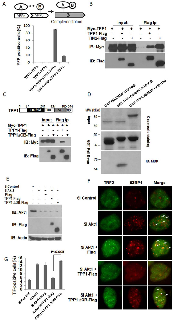
Figure 4. TPP1 can homodimerize through its OB fold.
(A) HTC75 cells co-expressing YFPn-tagged TPP1 with YFPc-tagged TPP1 or TIN2 were examined by BiFC assays. The percentage of cells displaying fluorescence complementation was quantitated by flow cytometry. Error bars indicate standard error (n=3). (B) 293T cells transiently co-expressing Myc-tagged TPP1 with Flag-tagged TPP1 or TIN2 were immunoprecipitated with anti-Flag antibodies. The immunoprecipitates were western blotted as indicated. (C) 293T cells transiently co-expressing Myc-tagged TPP1 with Flag-tagged TPP1 or TPP1 OB-fold deletion mutant (TPP1ΔOB) were immunoprecipitated with anti-Flag antibodies. The immunoprecipitates were western blotted as indicated. (D) Bacterially purified GST-tagged TPP1 OB fold only mutant (TPP1 OB) was incubated with MBP-tagged TPP1 OB for GST pull-down assays. The precipitates were resolved by SDS-PAGE and visualized by Coomassie staining or western blotting. GST-tagged Raf-1 Ras-binding domain (RBD) and MBP-tagged FAM118B were used as controls. (E) HTC75 cells were transfected with siRNA oligos against Akt1 (siAKT1-1) in combination with Flag-tagged wildtype TPP1 or TPP1 OB fold deletion mutant (TPP1ΔOB), and then analyzed by Western blotting using the indicated antibodies. Actin was used as loading control. (F) Cells from (E) were examined by immunostaining using anti-53BP1 (red) and TRF2 (green) antibodies. Arrows indicate overlapping foci. (G) Quantification of data from (F). Only cells with >4 co-localized foci were scored. Error bars indicate s.e.m. (n=3). P-values were determined by the Student t-test.

