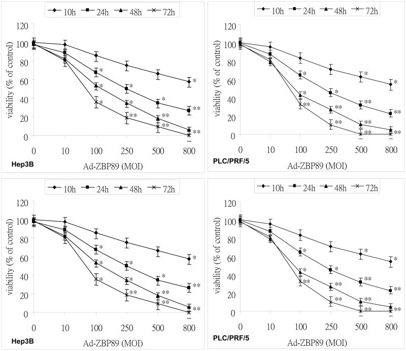
Figure 1. Celldeath induced by ZBP-89.
Cell death, reflected by the reduction of cell viability, was determined by MTT assay. Hep3B, PLC/PRF/5, SK-Hep-1 and Chang cells were culture in 96 well plates and infected with Ad-ZBP-89 at different concentrations. Cells infected with empty Ad5 vector were set up as control for each experiment. The infected cells were incubated for 10, 24, 48 and 72 huntil the MTT assay was performed. The viability of the cells with treatment was expressed as a relative value to that of the control. Data were reported as the mean±SD of three separate experiments. The significant difference in cell viability was observed between the cells without Ad-ZBP-89 treatment and those with Ad-ZBP-89 at indicated time points (*p<0.05 and **p<0.01).

