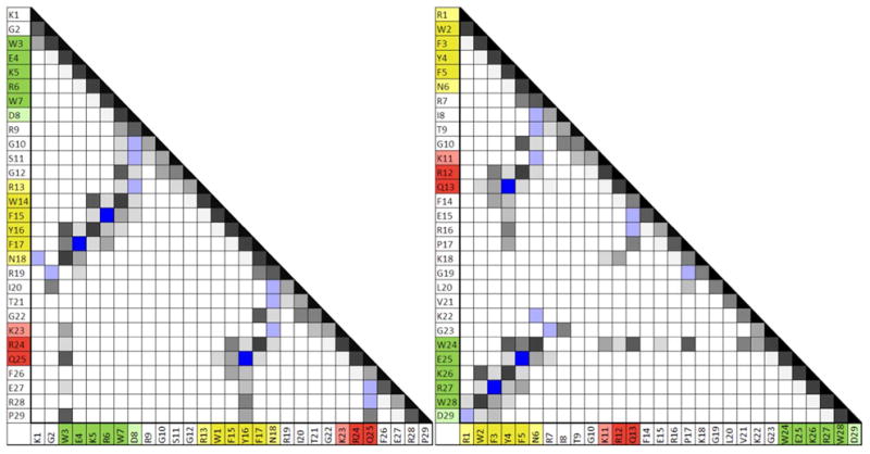
Figure 6.

Contact plots for the 29 residue standard-topology WW-domain mutant (left) and its circular permutant (right). Contacts with more and/or more intense NOEs are shown in darker shades of gray. H-bonding interactions are shown in blue. (These are evidenced by chemical shift deviations as well as NOEs. Deeper blue = two cross-strand backbone H-bonds are present.) The three strands of the core WW domain structure are color-coded: strand 1 is green, strand 2 is yellow, and strand 3 is red; the 1, 2, 3 numbering is based on the standard topology (see figure 1). Note the greater prevalence of much longer range interactions in the circular permutant.
