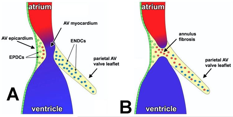
Figure 3.
Schematic representation of the contribution of the AV epicardium and the epicardially-derived cells to the development of the AV junction. After formation of the epicardial epithelium (green), epiEMT generates a population of AV-EPDCs (green cells in panel A) that, as far as their gene expression profile is concerned, are still very similar to the epicardium itself. However, when the AV-EPDCs migrate further into the AV sulcus and approach the AV myocardium (red cells), the molecular profile of the AV-EPDCs changes drastically as the expression of genes characteristically found in the mesenchyme of the annulus fibrosus (e.g., MMP2) and the AV cushions (e.g., Sox9) is upregulated. These “differentiated” AV-EPDCs (red cells in panel A) then penetrate the AV myocardium to form the annulus fibrosus (panel B) and migrate into the parietal AV valve leaflets where they intermingle with the endocardially-derived mesenchymal cells (ENDCs; blue cells in panels A and B).

