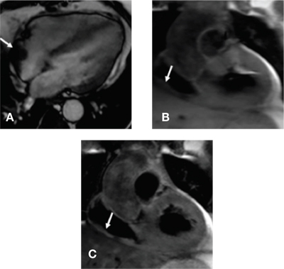
Figure 1.
Cardiac MRI: Image A is four-chamber steady state free precision showing mass in the posterior aspect of the right atrium that appears to be broad-based with extension into the right atrium (white arrow). Images B and C are T1 and T2 turbo-spin echo images showing the mass in the right atrium has intermediate intensity (white arrow).