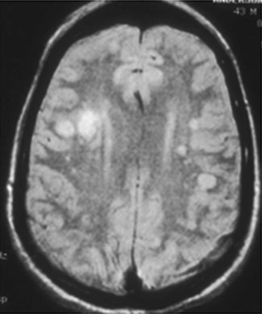
Fig. 2.
Brain magnetic resonance image (MRI) of a patient with varicella zoster virus multifocal vasculopathy. Proton-density brain MRI scan shows multiple areas of infarction, particularly involving gray–white matter junctions, in both hemispheres.(With permission from Gilden DH, Mahalingam R, Cohrs RJ, Kleinschmidt-DeMasters BK, Forghani B. The protean manifestations of varicella-zoster vasculopathy. J Neurovirol 2002;8(Suppl. 2):75–9) [53]

