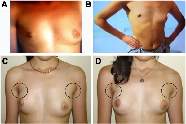
Figure 1.

Chest images from the twin girls. A and C) First twin. B and D) Second twin. A and B) Breast asymmetry, depression of the anterior chest wall indicating pectoral muscle hypoplasia, cranially located nipple, and hypoplastic areola are visible on the right side of both twin girls before surgery. C and D) Slight asymmetry and pectoral muscle hypoplasia are still perceivable, more evident in the first twin (left), two years after surgery.
