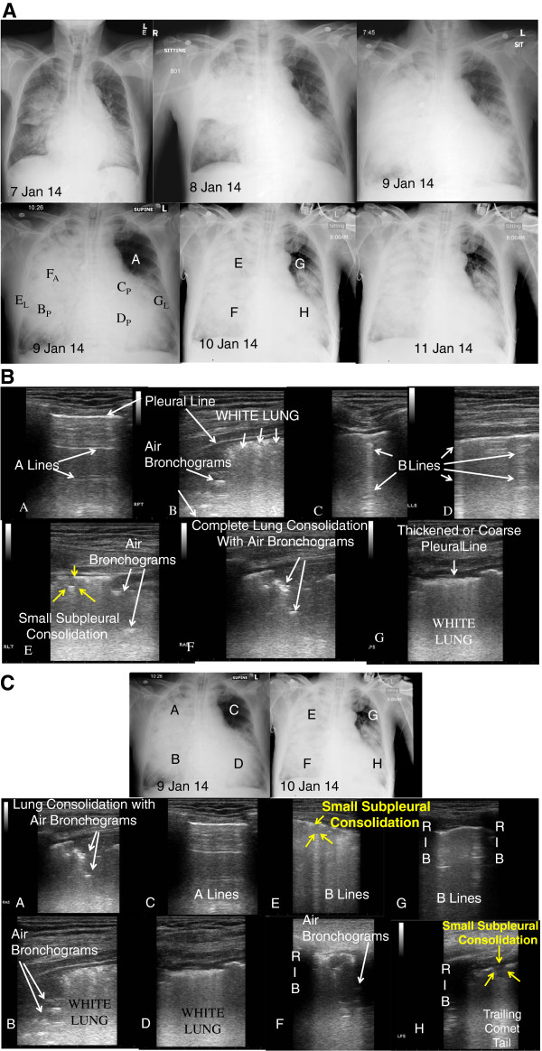
Figure 2.

Radiography and ultrasonography for case 2. (A) Chest X-rays showing progression over course of illness. CXR Letters correspond to panel letters in Figure 2B and 2C. Subscript A - anterior, P - posterior, and L- lateral. (B) Lung ultrasound images correlated to chest X-ray 9 Jan 14 in Fig. 2A. Panels A-G. (C) Correlated lung ultrasound images with chest X-rays over 2 days showing disease progression, particularly the left upper lobe (panels C and G). Paired panels by anatomic area: A + E; B + F; C + G; and D + H.
