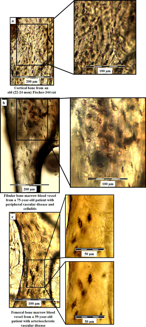
Figure 3. Osteocyte lacunae on rat cortical bone and bone marrow blood vessels isolated from human fibular and femoral diaphyses.
Light microscopic images of cortical bone from the femoral diaphysis of an old male Fischer-344 rat (a) and fibular and femoral bone marrow blood vessels from human patients with peripheral vascular disease and cellulitis (b) and arteriosclerotic vascular disease (c). Higher magnifications illustrate the similarities in osteocyte lacunar morphology between the cortical bone and ossified blood vessels.

