
Fig.1. Environmental challenges activate HSF1 signaling in the embryonic cerebral cortex.
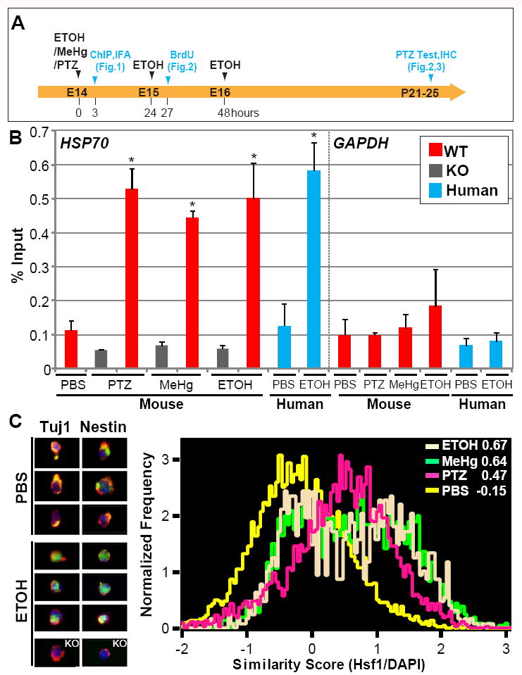
(A) Experimental scheme for Figs.1-3. (B) Chromatin immunoprecipitation analysis showing that the association of HSF1 with the HSP70 promoter is increased in embryonic mouse and human cortices after exposure to the indicated substrates. Occupancy of Hsp70 and Gapdh promoters was analyzed 3 hours after the challenge exposure. Quantification of the band intensities compared with input is shown. “WT” and “KO” indicate wild-type and Hsf1 KO mouse samples, respectively. *p < 0.005 by Student’s t-test compared with controls (n = 4 samples per condition, experiments were repeated at least two times for each sample). (C) Subcellular localization analysis of Hsf1 shows nuclear translocation of Hsf1 by prenatal exposure to indicated challenges. Left panels show representative flow cytometry images of co-immunostaining for Hsf1 (green) and Tuj1 or Nestin (red) with DAPI nuclear counter-stain (blue) in the cortical cells dissected from control (PBS)- and ETOH-exposed embryos. The cells from Hsf1 KO mouse embryonic cortices did not show Hsf1 labeling, confirming the specificity of labeling by the Hsf1 antibody (images at the bottom). Subcellular localization was analyzed using Imaging flow cytometry (right). Numbers at the right top corner are similarity scores indicating the extent to which the fluorescent signals of Hsf1 labeling coincided with nuclear DAPI staining in each cell. Analysis for each condition included more than 100,000 cells obtained from the embryos from multiple litters. See also Figure S1.