
Fig.4. Increase of apoptotic cell death in the cortex of Hsf1 KO embryos exposed to challenges.
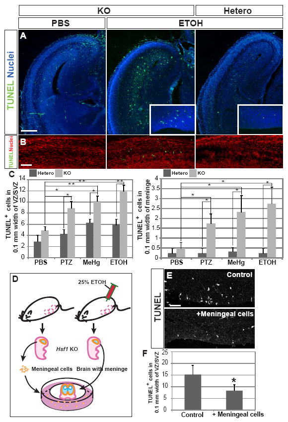
(A) TUNEL staining at E16 in cortical slices from embryos with indicated genotypes and prenatal exposure. An increase in apoptosis in the cortex of Hsf1 KO embryos exposed to ETOH is evident, particularly in the VZ/SVZ (insets). Bar = 0.5 mm. (B) TUNEL staining in meninges of the E16 Hsf1 KO embryos exposed to PBS or ETOH. ETOH-exposed Hsf1 KO embryos show increased apoptosis. Bar = 50 μm. (C) Quantification of TUNEL+ cells in the VZ/SVZ (left) and in the meninge in the cortices of Hsf1 mice exposed to indicated substrates. *p < 0.05; **p < 0.0001 by Student’s t-test. n = 8 (left) or 4 (right) embryos from more than 3 dams for each experimental condition. (D) Scheme for the preparation of in vitro slice culture with meningeal cells (see the details in Experimental Procedures). Slices of cerebral cortex and surrounding tissues were prepared from the Hsf1 KO embryos exposed to ETOH at E14 and 15, 3 hours after the last exposure to ETOH at E15. Fig.S3F demonstrates the increase in apoptosis in the meningeal cells at this time. The slices were cultured on filter inserts in the plates on which meningeal cells collected from other Hsf1 KO embryos (without challenge exposure) were plated. (E) Compared with control cultures (without meningeal cells, top), the culture with meningeal cells from embryos unexposed to challenge (bottom) showed significantly decrease in TUNEL+ cells in the VZ/SVZ. Bar = 50 μm. (F) Quantification of TUNEL+ cells in the VZ/SVZ. *p = 0.027 compared with the control culture (without meningeal cells) by Student’s t-test. n = 4 samples from 2 independent experiments per condition. See also Figure S3.