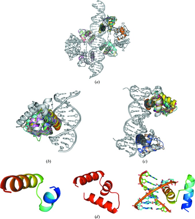
Figure 7.
(a) Group of HTH-type protein test cases used and search models. Target 2ISZ (space group P1) consists of four HTH fragments coordinated to a rather long DNA double strand. HTH-type fragment subsets are aligned with the target (shown in rainbow). Helix–turn–helix proteins are shown in grey and HTH-type search fragments are shown in rainbow. (b) HTH-type protein at 2.0 Å resolution with one HTH-type binding motif (PDB entry 3PVV; space group P3221) used as the target structure. All HTH-type fragment subsets are also aligned with the HTH target (rainbow). (c) HTH-type protein at 1.7 Å resolution with two HTH-type binding motifs (PDB entry 3RKQ; space group P65) used as the target structure. (d) Left, HTH-type search fragments (rainbow); middle, three-helix bundle HTH starting fragment (red); right, DNA including HTH-type fragment subsets as a search fragment (rainbow).

