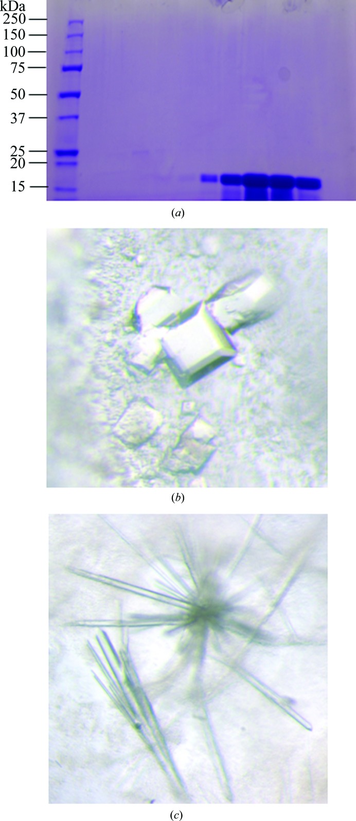
Figure 1.
Purification and crystallization of CypD. (a) SDS–PAGE of purified CypD protein stained with Coomassie Brilliant Blue. Lane 1, 7.5 µg CypD protein; lane 2, 15 µg CypD protein; lane 3, 30 µg CypD protein. (b) CypD-t crystals obtained from ProPlex condition D1. (c) CypD-o cystals obtained from Wizard 3–4 condition H4.

