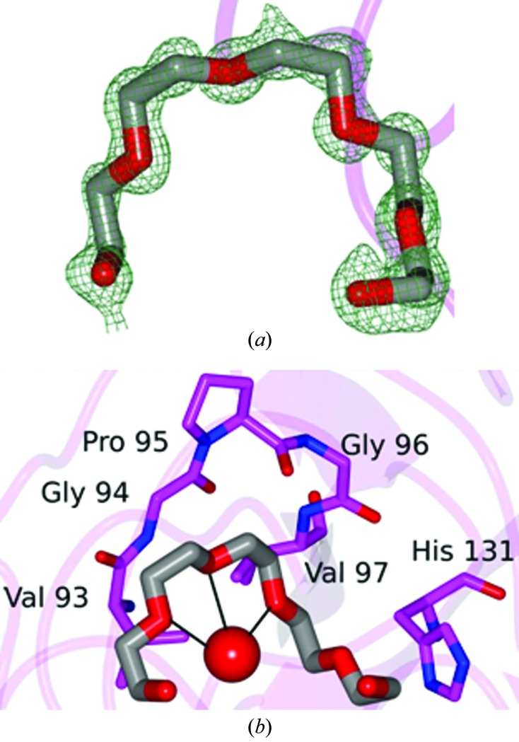
Figure 3.

PEG 400 molecule bound to CypD-t. (a) F o − F c electron-density map contoured at 3σ. (b) Hydrophobic residues that are in proximity (<4 Å) to the PEG 400 molecule. A water molecule (red sphere) is within hydrogen-bonding distance as indicated by the black lines.
