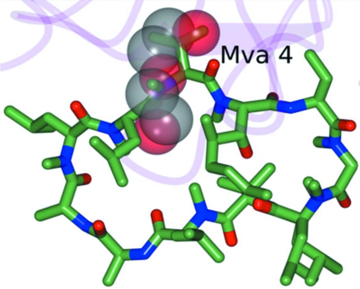
Figure 5.

Superposition of the partially disordered PEG 400 molecule in CypD-t (transparent spheres) with cyclosporine A (green) bound to CypD as observed in PDB entry 2z6w (Kajitani et al., 2008 ▶). The partially disordered PEG 400 molecule occupies a position similar to Mva4 of cyclosporine A as indicated. The r.m.s.d. between CypD-t and 2z6w is 0.33 Å between Cα atoms (164 residues).
