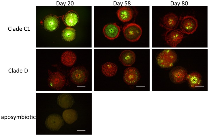
Figure 3. Fluorescence microscopic images of Acropora tenuis polyps associated with clade C1 or D Symbiodinium algae, and aposymbiotic polyps 20, 58, and 80 days after inoculation.
Bright green fluorescence can be seen in the body walls and tentacles of polyps associated with clade C1 algae. On day 20, polyps associated with clade D algae show little green fluorescence, and aposymbiotic polyps have a pale fluorescence. Red fluorescence dots indicate the chlorophyll fluorescence of Symbiodinium algae. Polyps with clade C1 algae contain few Symbiodinium cells, although many cells are aggregated around the polyps on days 20 and 58 (Fig. 3), thought to be Symbiodinium algae released from the corals. Scale bar = 0.5 mm.

