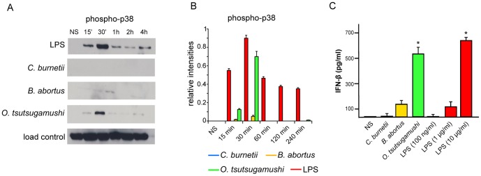
Figure 5. p38 phosphorylation and IFN-β release.

A, moDCs were stimulated with bacterial pathogens or E. coli LPS for different durations, ranging from 15 minutes to 4 hours, and immunoblotting was used to assess MAPK p38 phosphorylation. B, Densitometric scanning was used to quantify phosphorylation changes of p38 and the results are the mean ± SD of three experiments. C, moDCs were stimulated with bacterial pathogens or different concentrations of LPS for 16 hours. The release of IFN-β by moDCs was determined by ELISA. The results are expressed in pg/mL and represent the mean ± SD of three independent experiments.*p<0.03.
