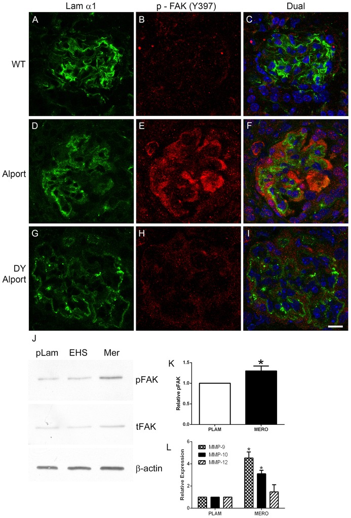
Figure 2. Laminin α2, but not laminin α1 activates FAK on podocytes in vivo and in vitro.
Panels A–C; 7 week old wild type glomerulus stained with antibodies specific for laminin 111 and pFAK397 show absence of pFAK immunostaining. Panels D–F; 7 week Alport glomerulus stained with antibodies specific for laminin α1 and pFAK397 pFAK immunostaining in podocytes adjacent to laminin α1-immunopositive GBM. Panels G-I show the same immunostaining as for D–F using Alport mice that do not express laminin α2 (the dy/dy muscular dystrophy mutation). Note the absence of pFAK397 immunostaining even though GBM is immunopositive for laminin α1. Panel J. Wild type podocytes were differentiated for 2 weeks and then plated on placental laminin, EHS laminin, or merosin for 15 hours. Extracts were prepared and analyzed by western blot for expression of pFAK397 and total FAK. β-actin was used as a loading control). Panel K shows quantitative analysis of pFAK397 relative to total FAK for several western blots. Panel L shows real time qRT-PCR results for transcripts endocing the indicated MMPs, demonstrating significantly elevated expression of MMP-9 and MMP-10 for cells cultured on merosin (MERO) relative to cells cultured on placental laminin (PLAM). Scale bar = 10 µm.

