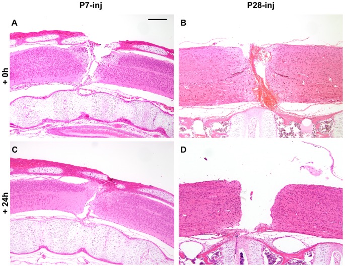
Figure 1. Monodelphis domestica spinal cords injured at P7 or P28.
Longitudinal sections (hematoxylin & eosin staining) of spinal cords injured at P7 or P28 shown immediately after complete spinal transection at T10 (A, B) or 24 hours later (C, D). Note obvious bleeding into the injury site at P28 (B), which was more pronounced than at P7 (A) One day after transection (+24 h) the gap between severed ends of the cord was larger in P28 injured animals (D) than in P7 injured animals (C). Rostral end is to the left, caudal to the right, dorsal is uppermost. Scale bar is 500 µm.

