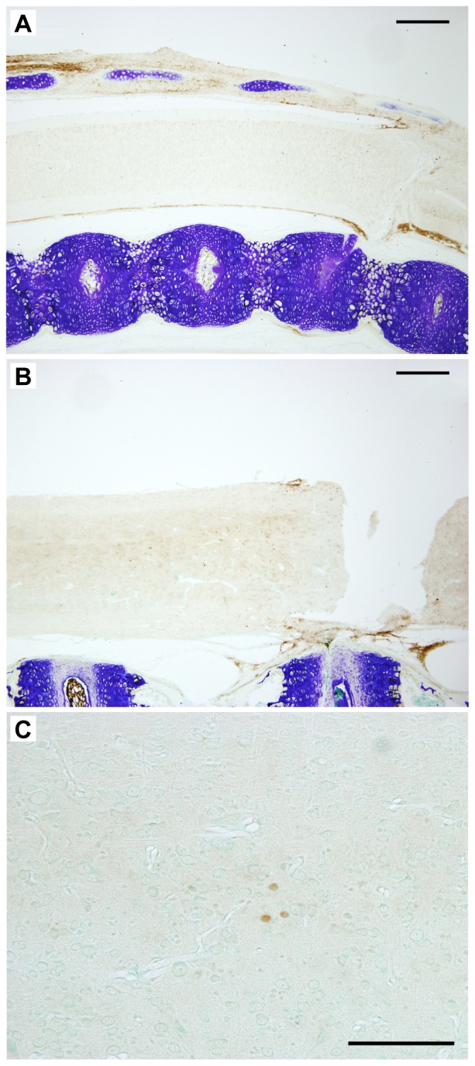
Figure 4. Interleukin-1β in Monodelphis spinal cord 24 hours after a complete transection at P7 or P28.

In the segment of the cord rostral to the site of injury Il-1β was detected using cross-reacting antibodies to the human cytokine. Note strong immunopositive signal in the tissue surrounding the cords at P7-injured (A) and P28 (B) but lack of significant staining within the spinal tissue especially at P7 (A). One day following injury at P28 a few immunopositive cells with the general morphology of monocytes were detected, especially in segments of the cord more rostral to the injury (C). Scale bars A, B = 500 µm, C = 100 µm.
