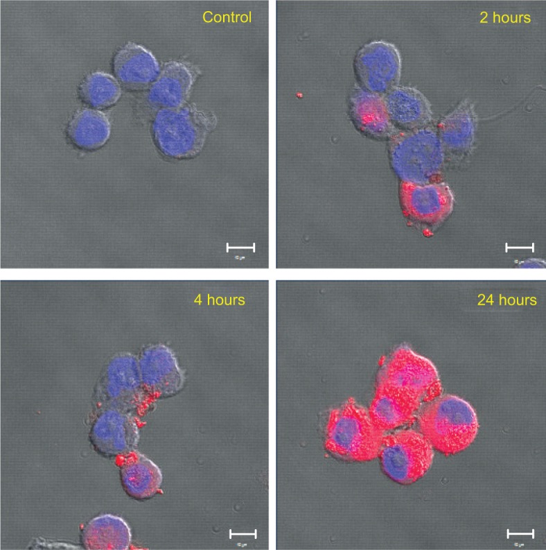
Figure 6.

Cellular uptake of PS-PDLLA NPs in PC-3 cells.
Notes: Cellular uptake and distribution of DiI-loaded NPs were observed by CLSM. DiI (1 μg/mL)-loaded NPs were incubated for 2, 4, and 24 hours. Red and blue colors indicate DiI and DAPI, respectively. Scale bar =10 μm.
Abbreviations: CLSM, confocal laser scanning microscopy; DAPI, 4′,6-diamidino-2-phenylindole; DiI, 1,1′-dioctadecyl-3,3,3′,3′-tetramethylindocarbocyanine perchlorate; NP, nanoparticle; PS-PDLLA, poly(styrene)-b-poly(DL-lactide).
