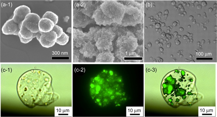
Figure 2.
(a) Typical scanning electron microscopy image of mesoporous silica nanoparticles (MSNs) used in this study, with (a-1) spherical morphology and (a-2) bulk-like morphology. (b) A typical optical microscopy image of alginate microspheres. (c) Fluorescent images of a fluorescein isothiocyanate-MSN-encapsulated alginate microsphere: (c-1) DIC image, (c-2) green fluorescent image from fluorescein isothiocyanate-adsorbed MSNs, and (c-3) merged image.
Abbreviation: DIC, differential interference contrast microscopy.

