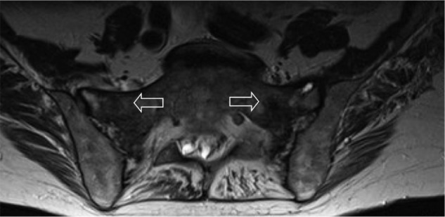
Figure 2.

Axial T1-weighted MRI image of sacrum.
Note: Patchy, low-intensity edema evident (arrows).
Abbreviation: MRI, magnetic resonance imaging.

Axial T1-weighted MRI image of sacrum.
Note: Patchy, low-intensity edema evident (arrows).
Abbreviation: MRI, magnetic resonance imaging.