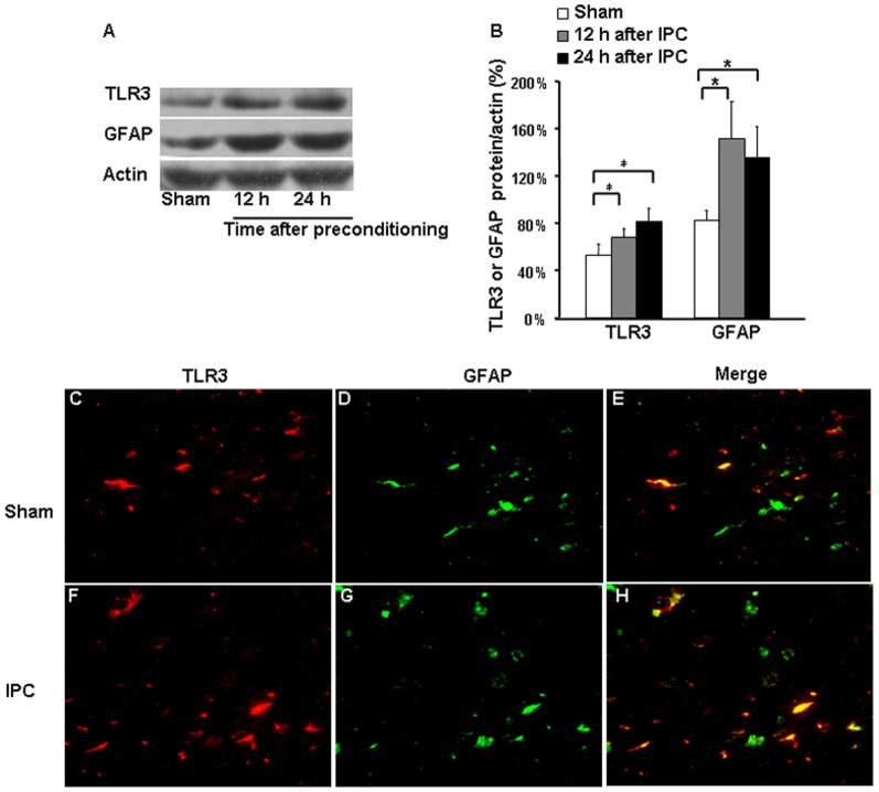
Figure 2. TLR3 and GFAP expression in cerebral cortex after ischemic preconditioning (IPC).
(A and B) Western blotting showed that protein expression of Toll-like receptor 3 (TLR3) and glial fibrillary acidic protein (GFAP) was elevated at 12 h and 24 h after IPC compared with that in sham-operated controls. (C and F) TLR3 immunoreactivity. (D and G) GFAP immunoreactivity. (E and H) Merged images show that co-localization of TLR3 and GFAP was increased at 24 h after IPC. Values are expressed as mean ± SD; n = 3 per group; *p<0.05. Magnification in C-H: 200x.

