
Figure 3.
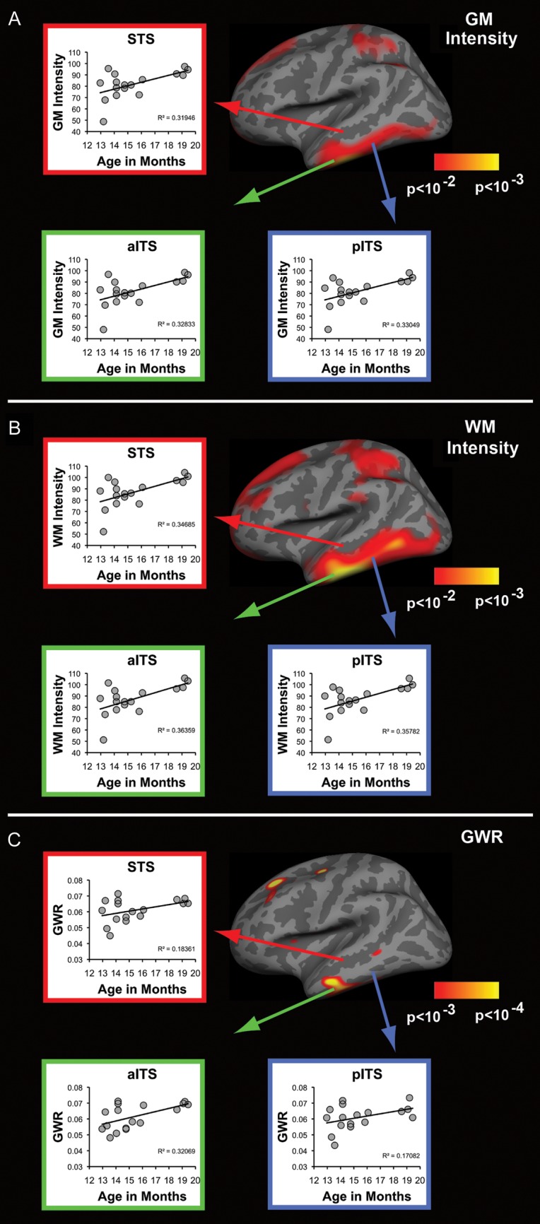
Tissue intensity and contrast significantly change with age within regions shown to generate lexico-semantic activity in infants and adults. (A–C) Results of correlations performed between age and all 3 tissue signal intensity measures for 3 left lateral temporal ROIs. (A,B) WM and GM signal intensity significantly increases with age for all 3 ROIs. (C) Contrast significantly increases with age within left anterior inferior temporal sulcus, with trends observed in the superior temporal sulcus and posterior inferior temporal areas. STS, superior temporal sulcus; aITS, anterior inferior temporal sulcus; p, posterior. GLM maps from Figure 2 are reproduced for convenience.