
Figure 8.
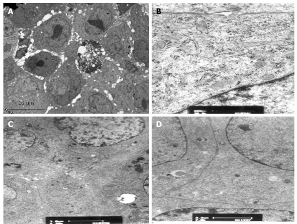
Transmission electron microscopy of alginate/chitosan-encapsulated cells in plasma before testing (× 12000). A: Low-power view of a single spheroid with an alginate bead shows cohesive colonies of cuboidal cells; B: High-power view showing cytoplasmic organelles, rough endoplasmic reticulum (RER), glycogen granules and mitochondria; C: Extensive microvilli, bile canalicular formation, junctional complexes and desmosomes; D: Tight junction.