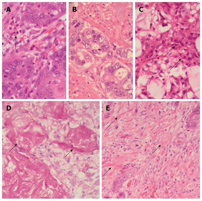
Figure 2.

Pathological features. A: Islands of malignant squamoid cells with surrounding stromal eosinophils (arrow) [hematoxylin and eosin (HE), × 480]; B: Glandular foci (HE, × 480); C: Foci of intermediate (arrows) and clear cells and of the cystically dilated mucinous component (HE, × 480); D: Southgate mucicarmine stain demonstrating the luminal (arrows) and intracytoplasmic mucin (southgate mucicarmine, × 480); E: Sclerotic stroma with keloid-like areas (arrows) (HE, × 480).
