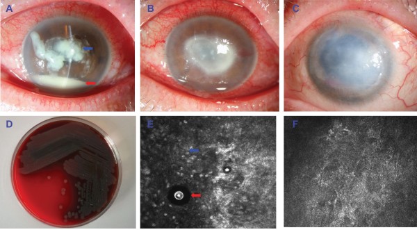
Figure 1.

An unusual keratitis case of coinfection with Acanthamoeba polyphaga and Pseudomonas aeruginosa. (A) Slit-lamp microscopic image of the left eye showed severe central corneal infiltrate (blue arrow) with intensive conjunctival injection. The anterior chamber had 20% hypopyon (black arrow). (B) After 1 week of treatment with topical antibiotics, a large central corneal ulcer with an underlying grayish-white, paracentral, ring-shaped stromal infiltrate was identified. (C) After 12 months of treatment with topical antibiotics, the left eye displayed no signs of inflammation, but there were some superficial blood vessels in the peripheral cornea and a large, central corneal scar obscuring the visual axis. (D) Microbiological cultures obtained from a superficial corneal swab showed the presence of Pseudomonas aeruginosa.I. (E)In vivo confocal microscopy examination showed the presence of oval to round, double-walled, highly refractile structures with a polygonal inner wall, varying 12–25 μm in size (red arrow), with infiltration of inflammatory cells (blue arrow). (F) The Acanthamoeba cysts could not be detected by IVCM examination after 12 months of treatment.
