
Figure 2.
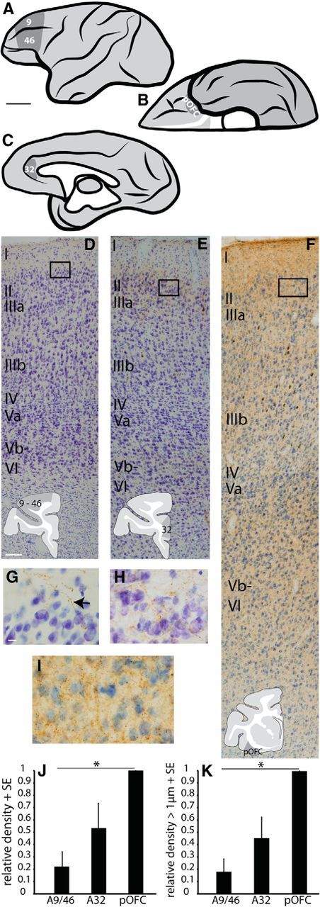
Amygdalar boutons were densest and largest in pOFC. A–C, Areas of study: lateral (A), orbital (B), and medial (C) views of a rhesus monkey brain show areas of interest in areas 9/46, OPro (pOFC), and A32 (ACC). Scale bar, 1 cm. D–F, Nissl-stained columns of cortex in areas 9/46 (D, A9/46); A32 (E); and pOFC (F, area OPro) labeled for amygdalar fibers (G–I, insets). Scale bar: (in D) D–F, 100 μm. G–I, Amygdalar fibers (arrow) at layer 1–2 border in A9/46 (G), A32 (H), and pOFC (I). Scale bar: (in G) G–I, 10 μm. J, Relative density of amygdalar boutons in A9/46, A32, and pOFC, normalized to the highest density in each case (pOFC). K, Relative density of large amygdalar boutons ≥1 μm in diameter in A9/46, A32, and pOFC, normalized to the highest density in each case (pOFC). Vertical lines indicate SE. *p ≤ 0.05.