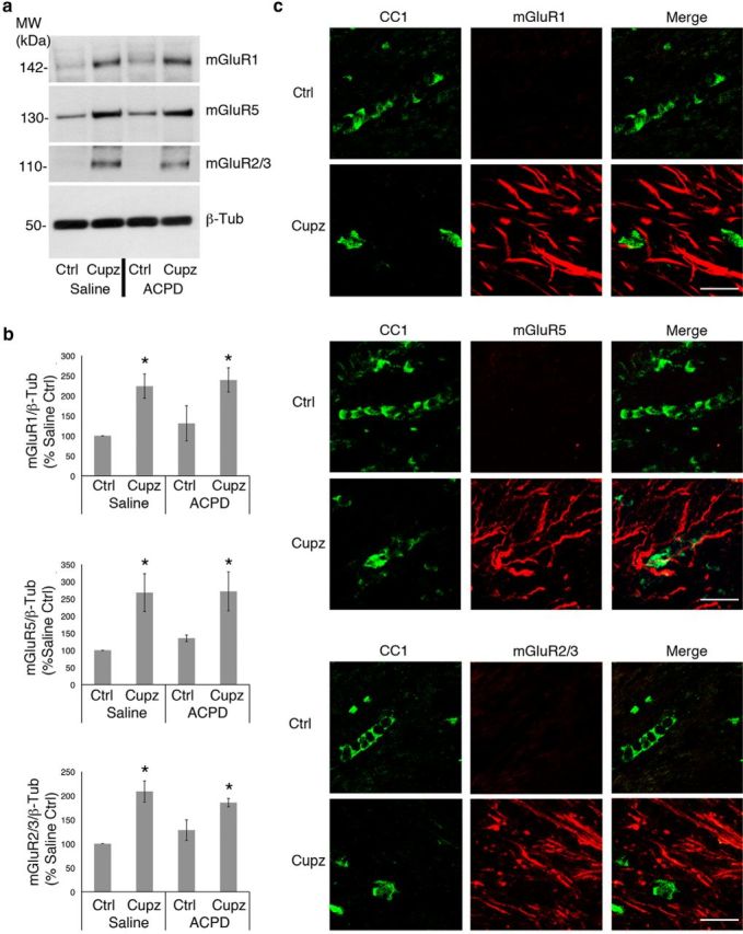
Figure 3.

Group I and Group II metabotropic glutamate receptor expression is increased in the cuprizone-lesioned corpus callosum. Wild-type mice were fed control or cuprizone-laden food for 4 weeks, then stereotaxically injected with either saline vehicle or ACPD. a, Western blots demonstrate mGluR1, mGluR5, and mGluR2/3 protein levels in the control and cuprizone-lesioned corpus callsoum. Beta-tubulin is shown as a loading control. b, Graphs represent a densitometric analysis of Western blots normalized to GAPDH and are presented as percentage saline-injected control. c, Immunohistochemical visualization of mGluRs and CC1+ oligodendrocytes. Western blot data analyzed by ANOVA; *significantly different from saline-injected control at p < 0.05. Each Western blot lane is from the corpus callosum of a single mouse within one experiment. Each experiment was repeated four times. Scale bar, 20 μm.
