
Figure 6.
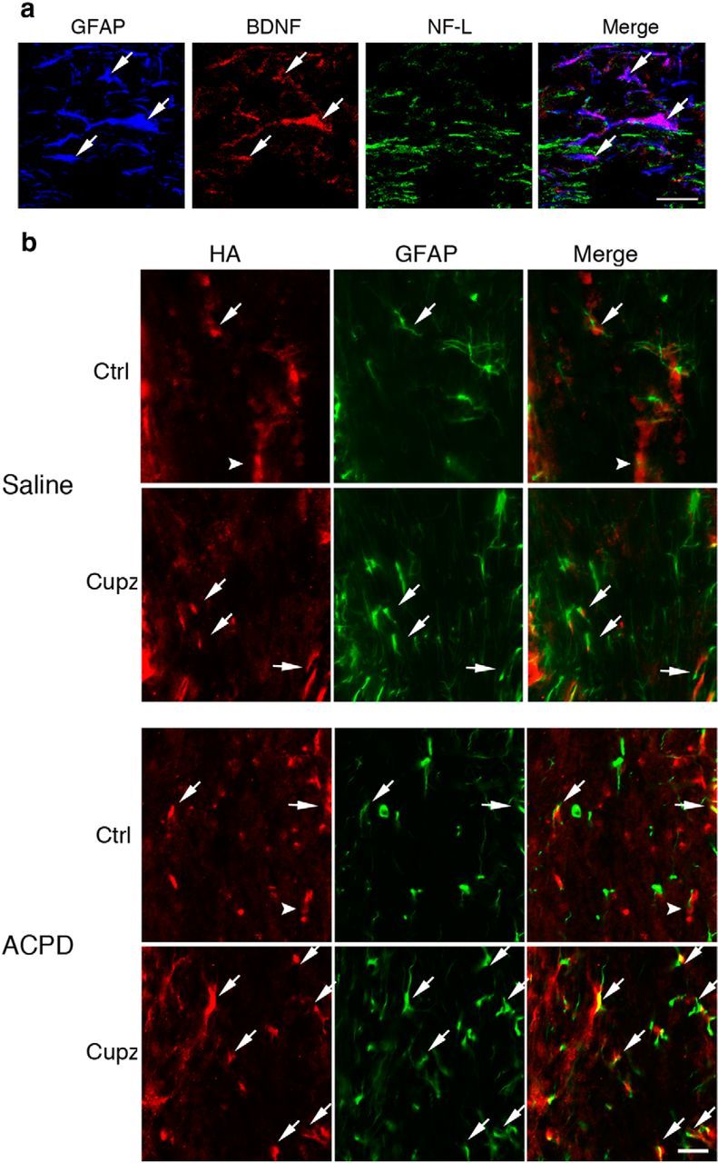
GFAP+ astrocytes express BDNF in the cuprizone-lesioned corpus callosum. Numbers of GFAP+ cells increase after ACPD. a, GFAP+ astrocytes in the cuprizone-lesioned corpus callosum colocalize with BDNF immunoreactivity. NF-L+ axonal tracts are not reactive for BDNF. b, HA-BDNF mice fed control or cuprizone feed for 4 weeks exhibit GFAP and HA immunoreactivity in only a small subset of the GFAP+ cells (arrows) in control mice or those fed cuprizone, whereas HA is predominantly found in non-GFAP+ cells (arrowhead) in controls. After cuprizone, the HA+ non-GFAP+ cells are greatly reduced. When cuprizone-treated mice are injected with ACPD, there is an increase in HA immunoreactivity associated with GFAP+ cells. These are representative images seen in three independent experiments. Scale bar, 20 μm.