
Figure 11.
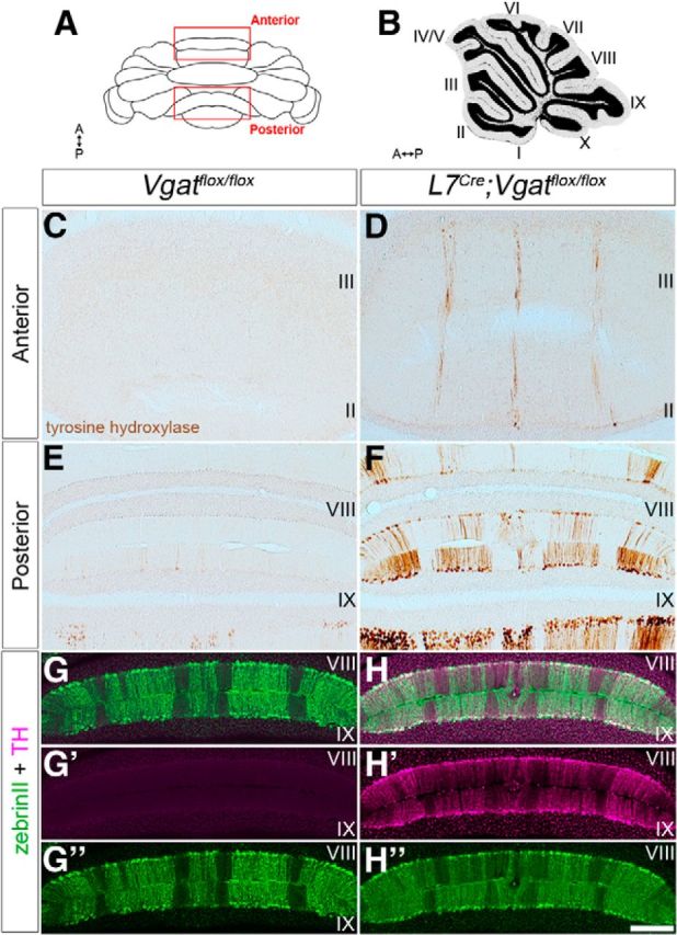
L7Cre;Vgatflox/flox mutant Purkinje cells express ectopic tyrosine hydroxylase (TH). A, Schematic diagram of the cerebellum shown in whole mount. A, anterior; P, posterior. B, Sagittal schematic of the cerebellum. The lobules are numbered with Roman numerals. C, Tyrosine hydroxylase expression was not detected in the anterior lobules of control mice. D, L7Cre;Vgatflox/flox mutant mice ectopically express tyrosine hydroxylase in narrow zebrinII-like zones. E, The posterior lobules of control mice have weak tyrosine hydroxylase expression in a subset of Purkinje cells. F, L7Cre;Vgatflox/flox mice heavily express tyrosine hydroxylase throughout the posterior cerebellum in a striking array of zones. G, H, ZebrinII and tyrosine hydroxylase merged images from control and mutant mice, respectively. G′, H′, Tyrosine hydroxylase expression in control and L7Cre;Vgatflox/flox mutant mice, respectively. G″, H″, ZebrinII expression in control and L7Cre;Vgatflox/flox mutant mice, respectively. Scale bar, 200 μm.