Figure 5.

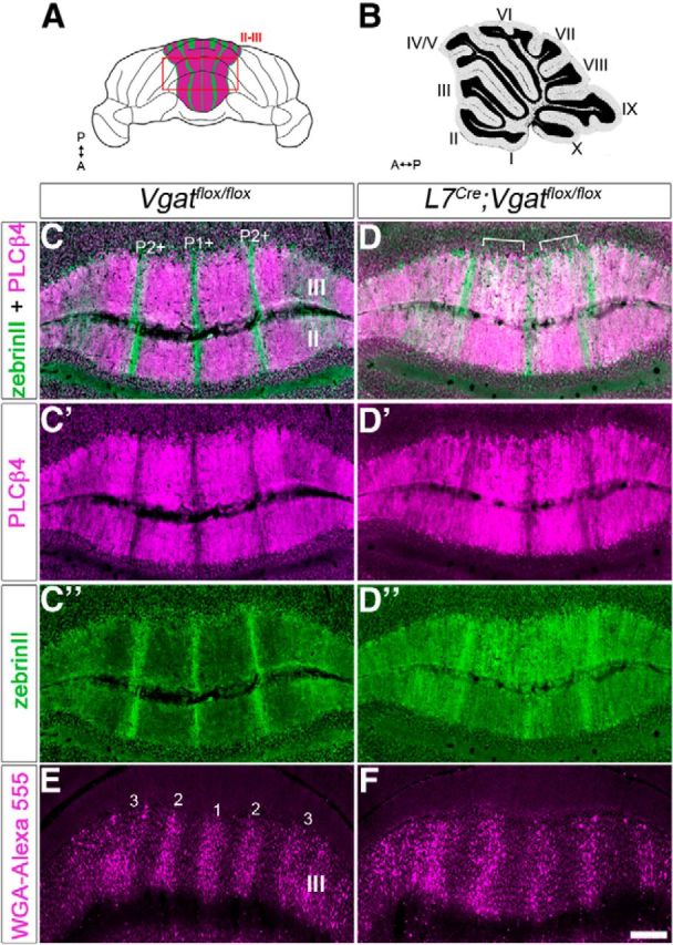
Zonal organization is altered in L7Cre;Vgatflox/flox mice (anterior lobules). A, Schematic whole-mount diagram of the cerebellum showing zebrinII and PLCβ4 expression in the anterior lobules. A, anterior; P, posterior. B, Sagittal schematic of the cerebellum. C, Coronal section cut through a Vgatflox/flox cerebellum showing zones in the anterior lobules II–III. ZebrinII-positive zones are marked as P1+ and P2+. D, Coronal section cut through an L7Cre;Vgatflox/flox cerebellum showing poor zonal boundaries. “White” staining represents overlapping zebrinII/ PLCβ4 expression (marked with brackets). C′, C″, D′, D″, Separated panels from C and D. E, Coronal section of a Vgatflox/flox cerebellum showing the zonal organization of spinocerebellar mossy fiber inputs in the anterior lobules. Mossy fiber terminal bands are numbered. F, Coronal section showing spinocerebellar zones in an L7Cre;Vgatflox/flox mutant mouse. Scale bar, 250 μm.
