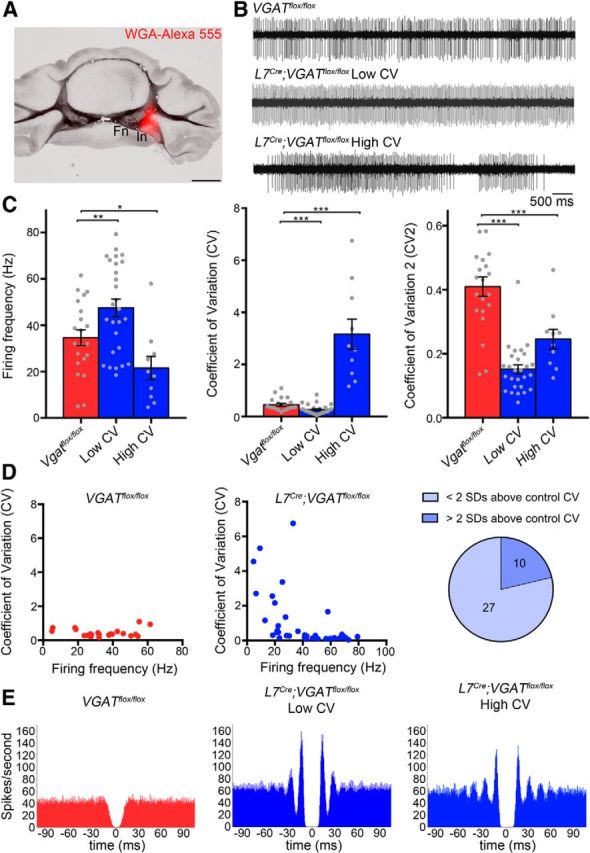
Figure 9.

L7Cre;Vgatflox/flox mutant mice exhibit abnormal firing of cerebellar nuclei neurons. A, An example of a cerebellar nuclei recording site marked by tracer injection. B, Sample raw in vivo electrophysiology traces from control and mutant cerebellar nuclei neurons. C, Quantification of firing frequency, CV and CV2 of control and mutant cerebellar nuclei neurons. D, Correlation between CV and firing frequency. The pie graph shows the percentage of L7Cre;Vgatflox/flox cells either less than or greater than 2 SDs above the control CV. E, Autocorrelograms of control and mutant nuclear cell spike trains reveal a flat autocorrelation histogram for control mice but multiple, equidistant side peaks with the primary peak centered at ∼15 ms for mutant mice; *p < 0.05, **p < 0.01, and ***p < 0.001.
