
Figure 7.
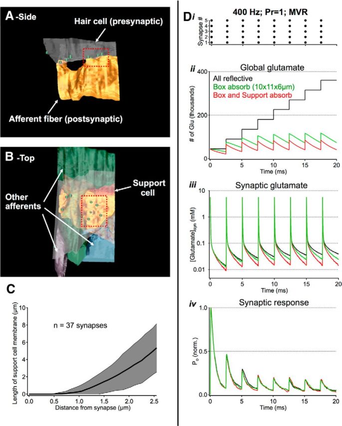
Active transport does little to shape rapid synaptic signaling. A, Zoomed out view of reconstruction used for synaptic simulations in Figures 4 and 6–8. B, The reconstructed environment included three other afferent fibers and one support cell (red). C, 2D analysis of individual micrographs indicates that most support cell membranes are >1 μm from individual synapses, limiting their ability to shape fast synaptic signaling. D, Exploration of active uptake during high-frequency release. i, Stimulus: 400 Hz release from all 5 synapses in the cluster, 9000 glutamate molecules released per event. ii–iv, A large absorptive bounding box (10 × 11 × 6 μm) helped to speed simulation time while making minimal contributions to local synaptic signaling. Contributions from support cells were tested by making their membranes absorptive to glutamate. This manipulation helped reduce global glutamate concentrations but had negligible effects on fast synaptic signaling.