Figure 3A.

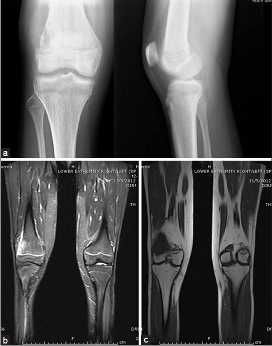
X-ray anteroposterior and lateral views (a) and coronal MRI T2W (b) and T1W (c) (preoperative imaging) showing osteosarcoma distal femur

X-ray anteroposterior and lateral views (a) and coronal MRI T2W (b) and T1W (c) (preoperative imaging) showing osteosarcoma distal femur