Figure 1.

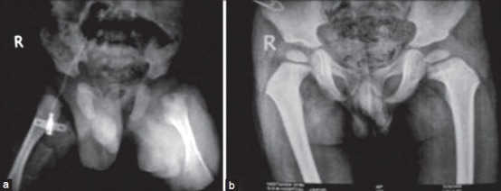
(Case 1) - X-ray pelvis both hip joints anteroposterior view showing (a) left hip subluxation, osteomyelitis upper end femur left side (b) followup at 2 years and 8 months. There is appearance of capital femoral epiphysis on both sides and coxa magna left side
