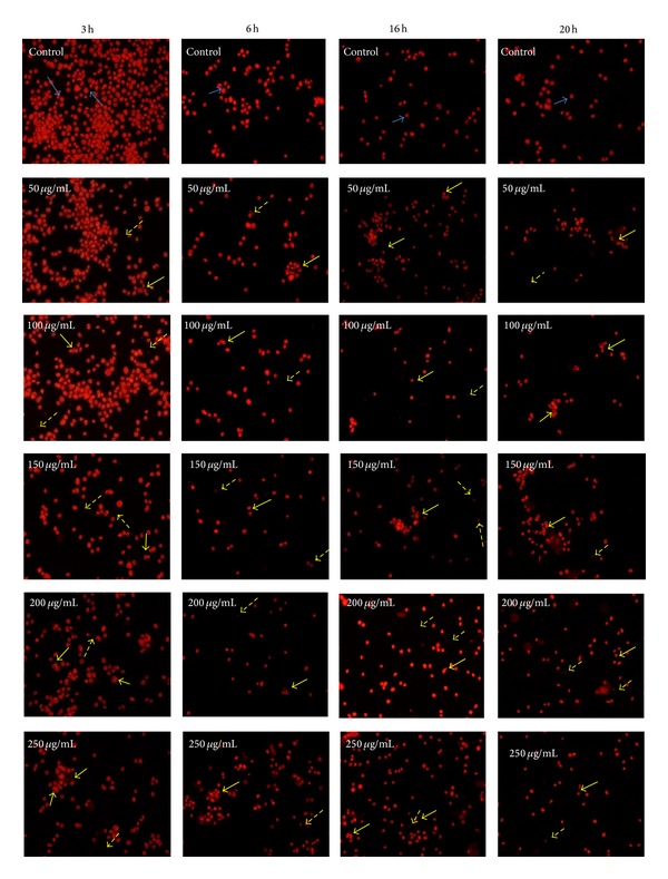
Figure 2.

PI staining of nuclei was done to examine morphological changes induced by D. formosum ethanolic extract at 50, 100, 150, 200, and 250 μg/mL concentrations and control under fluorescence microscope after treatment for 3 h, 6 h, 16 h, and 20 h, respectively [blue arrow shows live cells, yellow arrow represents apoptotic cells, and dotted yellow arrow shows presence of apoptotic bodies (late stage apoptosis)].
