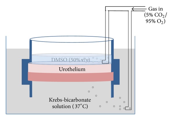
Figure 1.

Schematic figure of the incubation chamber. Full thickness sheets of bladder dome were sandwiched between two separated bathing solutions, with each containing gassed (5% CO2/95% O2) Krebs-bicarbonate (serosal) or DMSO (urothelial) solution at 37°C. Tissues were incubated with DMSO (50% v/v) applied to the luminal side only for 15 min before isolation of the various tissues for pharmacological analysis.
