Figure 1.

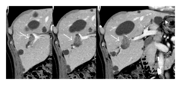
Representative images of intraductal papillary neoplasm of the bile duct on computed tomography. Localized bile duct dilatation and an intraductal mass are shown (arrows).

Representative images of intraductal papillary neoplasm of the bile duct on computed tomography. Localized bile duct dilatation and an intraductal mass are shown (arrows).