
Figure 7.
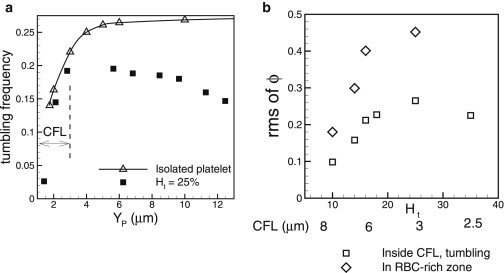
(a) Platelet tumbling frequency in dimensionless form as as a function of distance from the wall in whole blood (solid squares) at Ht = 25%, and for an isolated platelet (open triangles). The CFL thickness is 3 μm, as shown. (b) RMS of off-shear plane angle ϕ as a function of hematocrit. Shown here are the data for platelets flowing inside the RBC-rich zone (open diamonds), and for tumbling platelets flowing inside the CFL (open squares). The thickness of the CFL is shown below the lower axis.