Figure 7.

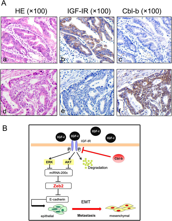
Representative images for IGF-IR and Cbl-b immunohistochemical staining in three serial sections from two cases. (A) (a, d) HE staining. The tumor is composed of atypical cells and irregularly shaped tubules. (b, f) IGF-IR and Cbl-b positive staining (+) in cell membrane and cytoplasm (in brown). (c, e) IGF-IR and Cbl-b negative staining (-). The original magnification is 100×. (B) Working model for Cbl-b in IGF-I-induced EMT. In gastric cancer cells, IGF-I binds to its receptor IGF-IR and activates Akt/ERK downstream signaling pathways. The activation of signal pathways up-regulates E-cadherin repressor ZEB2 and down-regulates miR-200c. There is an Akt/ERK-miR-200c-ZEB2 axis in the process. Ubiquitin ligase Cbl-b is required for sustaining the epithelial phenotype probably through targeting IGF-IR for degradation and further inhibiting Akt/ERK-miR-200c-ZEB2 axis in IGF-I-induced EMT.
