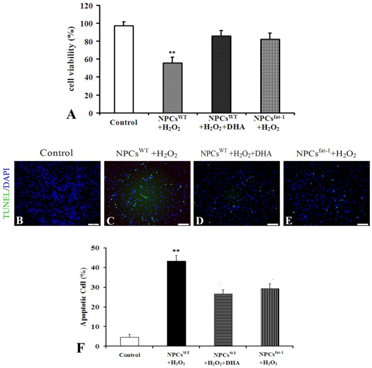
Figure 3.
NPCsfat-1 attenuated H2O2-mediated apoptosis. (A) The cell viability of NPCs was assessed after exposure to H2O2 for 6 h by WST-8 analysis. Each value represents the mean ± SD of three independent experiments (n = 3, ** p < 0.01 versus other groups); (B–E) Representative photomicrographs of TUNEL assay; (F) Quantitative analysis was carried out by measuring TUNEL-positive cells in each group. Figures were selected as representative data from three independent experiments. Cell apoptosis was significantly reduced in DHA-pretreated NPCsWT and NPCsfat-1. Each value represents the mean ± SD of three independent experiments (n = 3, ** p < 0.01 versus other groups). Scale bar: 75 μm.

