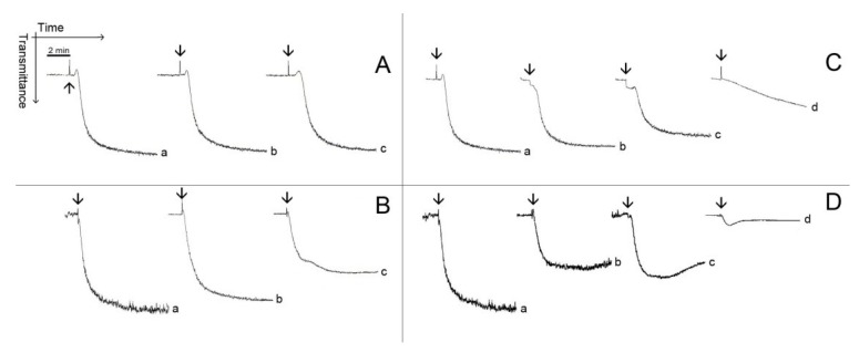
Figure 3.
Typical patterns of platelet aggregation with PRP. (A,B) PRP was incubated for 2 min at 37 °C while stirring with 0.69 mM (Line b) or with 1.38 mM (Line c) of pachydictyol A/isopachydictyol A, then 16 μg/mL of collagen (upper panel) or 15 μM of ADP (lower panel) were added to induce aggregation. (C,D) PRP was incubated as above, with 0.31 mM (Line b), 0.62 mM (Line c) or with 1.25 mM (Line d) of dichotomanol, and then collagen (upper panel) or ADP (lower panel) was added to the medium. For all panels, Lines a represent PRP incubated with 1% DMSO (v/v, final concentration). The arrows mark the addition of agonists.

