
Figure 2.
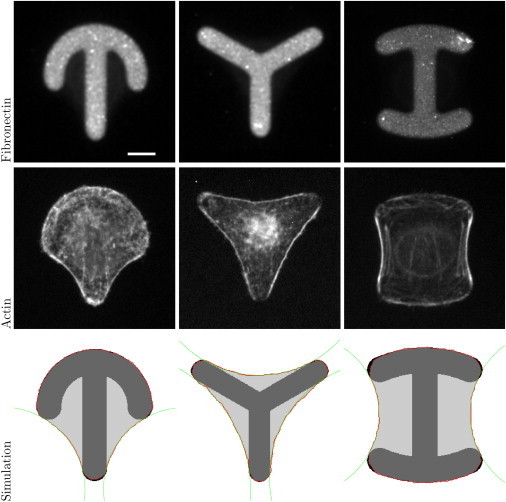
The top row shows experimental images for crossbow, Y, and H patterns coated with fibronectin. Scale bar, 10 μm. The middle row shows HeLa-cells stained for actin on those patterns. The bottom row shows shape predictions by the CPM with circles fitted to the free spanning arcs. Experimental images were kindly provided by Gintarė Garbenčiūtė and Vytaute Starkuviene-Erfle. To see this figure in color, go online.