
Figure 3.
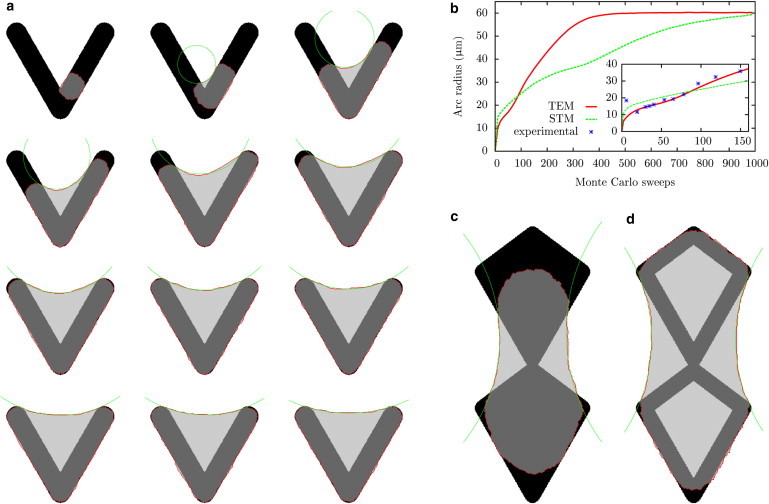
Cell shapes predicted by the CPM with surface tension σ = 0.6 nN/μm, simple tension λs = 10 nN, arc rigidity EA = 2000 nN, adhesive energy density W = 60 nN/μm, and cell target area A0 = 1700 μm2. (a) Cell spreading on a V-shaped pattern. The arms have an length of 46 μm and the final spanning distance and radius are d ≈ 33 μm, and R ≈ 61 nN/μm. A circle is fitted to the edge bundle. (b) Radius of the circle fitted to the edge as a function of Monte Carlo sweeps for cells described by the STM and TEM. The radius is averaged over 104 cells all starting to spread at the same position as in the previous figure. Here, each Monte Carlo sweep consists of 2 × 10⁴ attempts to invert one of the boundary lattice sites. For the simulation of the STM cells, a simple line tension of λs = 36.6 nN was used, resulting in the same final radius as for the TEM cells. (Inset) Initial spread phase and data taken from Théry et al. (6). (c) Final cell shape on an adhesive pattern that cannot be fully covered by the cell. Pattern has a width of 40 μm and a height of 96 μm. (d) Same as d, but with a hollow adhesive geometry. To see this figure in color, go online.