
Figure 6.
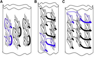
Models of amyloid fibrils built from three β-structured modules, each with three strands (arrows) and two turns. Lines between β-strands denote H-bonds. (A) β-meander model having three side-by-side β-sheets. Its width is between 2.7 and 3.7 nm and expected MPL is one molecule per cross section. In principle, β-meanders from the neighboring β-sheets can be axially displaced. (B) A β-solenoid model in a parallel conformation with estimated width of 1.8 to 3.7 nm and 2/3 molecule per fibril cross section. (C). A superpleated β-structure model in a parallel conformation with width of 2.9 to 3.7 nm and one molecule per fibril cross section.