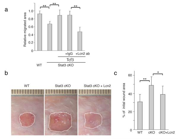
Figure 9. Lipocalin2 can rescue defective wound healing in Stat3 cKO mice.
(a) Migration assays were performed on Stat3+/fl:K14-cre (cont) or Stat3fl/fl:K14-cre (Stat3 cKO) keratinocytes that were transduced with control or tet-inducible Tcf3 treated with Dox together with anti-Lcn2 antibody or IgG isotype control. Graph quantifying the relative area migrated by keratinocytes normalized over area migrated by cells transduced with control vector without Dox. For each sample, over 30 non-overlapping fields were measured at each timepoint; and each experiment was repeated twice. Data are mean ± s.d. **p<0.001 (Student’s t-test).
(b) Images of skin wound sites taken 10 days post wounding. After full-thickness wounds of 1cm2 were created on the dorsal skins of wild-type and Stat3 cKO mice, 200μl of recombinant Lcn2 (2μg/ml) or vehicle were applied topically onto the wound sites every other day. Surface areas of the wounds were measured at the initial time point and 10 days post wounding.
(c) Graph quantifying the surface areas of the wounds as a percentage of the original wounds 10 days post wounding. wild-type (n=13) and Stat3 cKO (n=7). Data are mean ± s.d. *p<0.05 and **p<0.001 (Student’s t-test).

