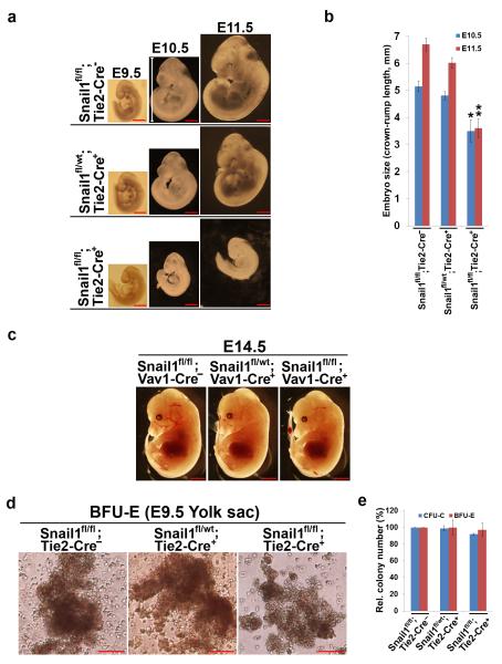
Figure 1. Endothelial-Specific Deletion of Snail1 Induces Embryonic Lethality.
(a) Gross examination of whole embryos at the indicated stages of development. Note the significantly reduced size of Snail1fl/fl;Tie2-Cre+ embryos at E10.5 and E11.5. Scale bar: 1 mm.
(b) Quantification of embryo size as indicated by crown-rump length (shown in [a]) (n=4 in each group). Data are presented as mean ± SEM. *, ** p < 0.05 and p < 0.01, respectively (ANOVA test).
(c) Gross examination of whole embryos from Vav1-Cre crosses at E14.5. No differences were observed between Snail1fl/fl;Vav1-Cre+ mutants and their control littermates in size, stage or overall appearance. Scale bars: 2 mm.
(d,e) In vitro differentiation analysis of yolk sac hematopoietic cells derived from E9.5 Snail1fl/fl;Tie2-Cre+ mutants and their control littermates. Representative photomicrographs of BFU-E colonies 7 d after plating are shown at left (d). Quantification of CFU-E and BFU-E colonies from yolk sacs (n=3 in each group) is shown to the right (e). Data are presented as mean ± SEM. Not significant, ANOVA. Scale bars: 50 μm.

