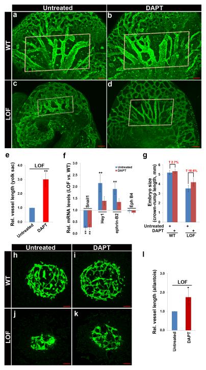
Figure 6. Administration of DAPT Partially Reverses Snail1 Deletion-Induced Vascular Defects.
(a-d) Confocal analysis of PECAM-1 stained whole-mounted untreated (a,c) or DAPT-treated embryos (b,d). Rectangled area highlights improved vascular remodeling in DAPT treated Snail1 LOF mutant embryos. Scale bar: 100 μm.
(e) Quantification of relative vessel length in yolk sacs from E10.5 untreated and DAPT treated Snail1 LOF embryos (n=4 each). Data are presented as mean ± SEM. *p < 0.05, **p < 0.01, Student’s t test.
(f) RT-qPCR analysis of ECs freshly isolated from untreated or DAPT treated E10.5 embryos (n=4 each). Data are presented as mean ± SEM. **p < 0.01, Student’s t test.
(g) Quantification of embryo size in a-d above.
(h-k) Allantoises dissected from WT (h,i) and Snail1 LOF mutant (j,k) embryos were cultured in the presence of vehicle (h,j) or 8 μM DAPT (i,k) for 24 h and ECs visualized by whole-mount PECAM-1 staining. Scale bar: 100 μm.
(l) Quantification of relative vessel length in allantoises (n=4 each). Data are presented as a mean ± SEM. **p < 0.05, Student’s t test.

